Amit Jain is Consultant and Head,
Amit Jain’s Institute of Diabetic Foot
and Wound Care, Brindhavvan
Areion Hospital, Bangalore;
Associate Professor, Department
of Surgery, Rajarajeswari Medical
College, Bangalore, India; Visiting
Consultant, MV centre of Diabetes,
Bangalore, India; Kishore Kumar
is Consultant, Amit Jain’s Institute
of Diabetic Foot and Wound Care,
Brindhavvan Areion Hospital,
Bangalore, India; Harish Kumar is
Consultants, Amit Jain’s Institute
of Diabetic Foot and Wound Care,
Brindhavvan Areion Hospital,
Bangalore, India; Suresh Kumar is
Consultants, Amit Jain’s Institute
of Diabetic Foot and Wound Care,
Brindhavvan Areion Hospital,
Bangalore, India.
Diabetic foot is one of the most common
complications in people with diabetes
mellitus and can lead to lower limb
amputation (Morshed, 2011). Amputation occurs
10–30 times more commonly in people with
diabetes when compared to general population
(Woodbury, 2015). It is known that 15% of
patients with diabetes are likely to develop a
foot ulcer in their lifetime (Yazdanpanah, 2015).
Around 56% of diabetic foot ulcers get infected
and many may end up in some form of lower
extremity amputation (Gibbons, 1984; Smith,
1987; Wu, 2015).
The diabetic foot on the whole is a classical
triad of neuropathy, infection and ischemia
(Pendsey, 2010; Jain, 2017). Early identification
of these can actually alter the disease process
and prevent amputation. This is best achieved
by screening to determine whether a disease
or condition is present. Screening is done in a
number of different ways, e.g. mass screening,
multiphasic screening, high-risk screening,
multipurpose screening and opportunistic
screening (Suryakantha, 2014; Park, 2015).
High-risk screening is screening of only a group
of population who are at a high risk of the disease
and not of the entire population. This is also
called Selective screening or Targeted screening
(Suryakantha, 2014; Park, 2015).
Screening of diabetic foot has to be considered
a preventive care strategy as it can prevent
amputation, which leads to debilitation with a
huge socioeconomic consequence.
Screening is quite different from a
diagnostic test (Park, 2015). It is believed
that screening should be inexpensive and
should require little physician or health care
professional time (Park, 2015).
The author believes that diabetic foot
evaluation can be either through screening
or through examination (Jain, 2017). There is
a well-known difference in both these types
of evaluation. Screening of foot is a quick
evaluation identifying those factors that lead to
risk of amputation. An examination of foot refers
to a detailed evaluation that can be a laborious
and time consuming method (Jain, 2017).
Diabetic foot screening can be undertaken by
any healthcare professional or ancillary staff
(Muzaini, 2017) whereas examination is often
done by a specialist.
Screening of diabetic foot is essential and the
author feels that it fulfills most criteria laid down
for screening (Park, 2015):
- It is an important health problem
- The pathway/natural history is adequately understood
- There is an asymptomatic stage. Example – neuropathy and peripheral vascular disease may remain asymptomatic
- There are tests that can detect the disease prior to onset of signs and symptoms
- Effective treatment can be instituted once disease is detected by screening
- There are also agreed policy and protocol on whom to treat
- Expected benefits of screening of diabetic foot by early detection exceed the risk and costs. There are many screening tools like In low’s screening tool, 60 seconds screening tool, etc (Jain, 2017) which are commonly used in different regions. Amit Jain’s triple assessment of foot is simple, safe and a rapid new screening
Giovinco NA, Millers JD (2015) A
Practical Update to Comprehensive
Screening in the High Risk Diabetic
Foot. Available at: http://www.
podiatrym.com/cme/CME215.pdf
(accessed 30.01.2018)
Jain AKC (2017) Amit Jain’s triple
assessment for foot in diabetes –
the simplest and the fastest new
screening tool in the world. IJMSCI
4(6): 3015–9
Jan M, Mattoo JA, Salroo NA et
al (2010) Triple assessment in
diagnosis of breast cancer in
Kashmir. Indian J Surg 72:92
Jayaprakash P, Bhansali A, Bhansali S
et al (2011) Validation of bedside
methods in evaluation of diabetic
peripheral neuropathy. Indian J
Med Res133(6): 645–9
Morshed GM, Mashahit Ma, shaheen
HA (2011) Simple screening
tests for peripheral neuropathy
as a prediction of diabetic foot
ulceration. FAOJ 4(11): 2
Muzaini AA, Baker N (2017) User’s
guide to diabetic foot screening.
The Diabetic Foot Journal Middle
East 3(2):14-21
Park K (2015) Screening for disease.
In: Park’s Textbook of Preventive
and Social Medicine (23rd edn).
Banarsidas Bhanot Publishers,
India
Pendsey SP (2010) Understanding
diabetic foot. Int J Diabetes Dev
Ctries 30(2): 75–9
Phulpoto JA, Gurbakhshani KM,
Shaikh A (2012) Role of bedside
methods in evaluation of diabetic
peripheral neuropathy. Rawal Med
J 37(2): 1–11
Smith D, Weinberger M, Katz B (1987)
A controlled trial to increase office
visits and reduce hospitalization in
diabetic patients. J General Int Med
2: 232–38
Suryakantha AH (2014) Epidemiology
of infectious disease. In:
Community Medicine with Recent
Advances (3rd edn). Jaypee
Publishers, India
Woodbury MG, Sibbald RG, Ostrow
B et al (2015) Tool for rapid easy
identification of high risk diabetic
foot: validation and clinical pilot
of the simplified 60 second
diabetic foot screening tool. Plos
One10(6):e0125578
Wu S (2015) Pressure Mitigation for
the Diabetic Foot Ulcer. Available
at: http://www.podiatrym.com/
pdf/2015/11/Wu1115web.pdf
(accessed 30.01.2018)
Yazdanpanah L, Nasiri M, Adarvishi
S (2015)Literature review on the
management of diabetic foot ulcer.
World J Diabetes 6(1): 37–53
ethod that was proposed recently from Indian
subcontinent (Jain, 2017). This screening is easy,
acceptable, repeatable and inexpensive (Park,
2015; Jain, 2017), that can be performed by any
health care professional in any part of the world
without difficulty. Amit Jain’s triple assessment
for diabetic foot addresses all the triad namely
neuropathy, infection and ischemia (Pendsey,
2010; Jain, 2017).
The author classified diabetic foot infections
in general into primary where infection occurs
directly into foot and secondary where the
infection occurs in pre-existing pathology like an
ulcer (Jain, 2017). Primary infections are usually
acute. Most of the Amit Jain’s type 1 diabetic foot
complications like abscess, cellulitis, necrotizing
fasciitis etc are primary diabetic foot infection
(Jain, 2017).
Amit Jain’s triple assessment of foot is a concept
that is derived from routine clinical examination
done in surgery (Das, 2008) and triple assessment
done in breast lump (Jan, 2010). This triple
assessment of foot has three components
namely: Look, Feel and Test which addresses all
the triad of diabetic foot very effectively namely
infection, ischemia and neuropathy.
In the Look component, an ulcer/infection is
identified. The parts of the foot needs to be seen
are dorsum of foot
[Figure 1], plantar surface
[Figure 2] and interdigital areas
[Figure 3]. In the Feel component, the dorsalis pedis/ posterior tibial artery is palpated
[Figure 4] to assess the blood flow to foot. An absent foot pulse should alert one for further assessment and investigation. In the Test component, neuropathy is detected by any of the following methods in isolation or preferably in combination and they include monofilament testing
[Figure 5], tuning fork, vibratip, biothesiometer, pin prick test, etc (Jayaprakash, 2011; Phulpoto, 2012; Arshad, 2016). The tuning fork, vibratip and biothesiometer are used to assess vibration sensation whereas the pinprick and monofilament is used to test touch sensation (Giovinco, 2015). One can use the above in combination to test both sensation and vibration and should check at least three to four sites. The common sites are plantar aspect of great toes, base of 1st, 3rd metatarsal, 5th metatarsal (Phulpoto, 2012; Giovinco, 2015; Arshad, 2016)
Advantages of the new screening tool
The advantages of Amit Jain’s triple assessment
are:
- It is a simple screening tool
- It is very practical
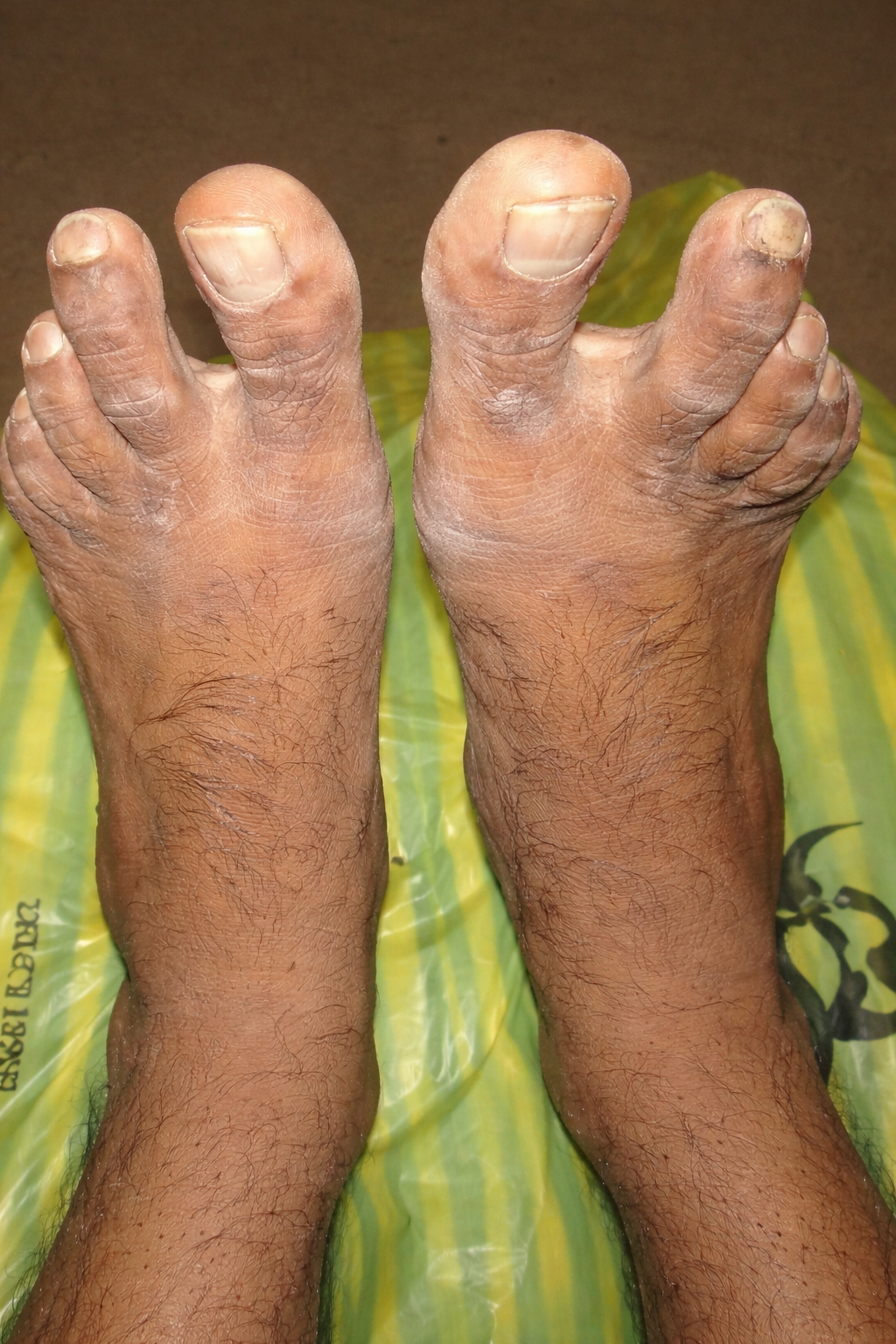
Figure 1. (left) The dorsum of the foot.
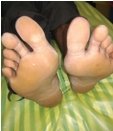
Figure 2.(right) The plantar aspect of foot.
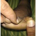
Figure 3. (left) The interdigital area.
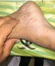
Figure 4. (right) Palpation of dorsalis pedis artery.
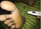
Figure 5. (above right). The palpation
of the posterior tibial artery.
- Easy to remember and perform
- It can be a good teaching tool
- Fulfills most criteria laid down for screening (Park, 2015)
- It addresses all the three components of diabetic foot effectively
- Any health care professional can use it
- It is serves as a good record of diabetic foot evaluation.
Sometimes Amit Jain’s single assessment
and double assessment for foot in diabetes is
performed in certain situations (Jain, 2017)
References
Arshad AR, Alvi KY (2016) Diagnostic accuracy of clinical
methods for detection of diabetic sensory neuropathy. J
Coll Phy Surg Pak26(5): 374–79
Das S (2008) A Manual of Clinical Surgery (12th edn). S Das
Publication, India
Gibbons G, Eliopoulos GM (1984) Infection of the
diabetic foot. In: Kozak GP Hoar CS Rowbotham JL, ed.
Management of